Symptoms and causes
Symptoms
The most common signs and symptoms of dermatomyositis include:
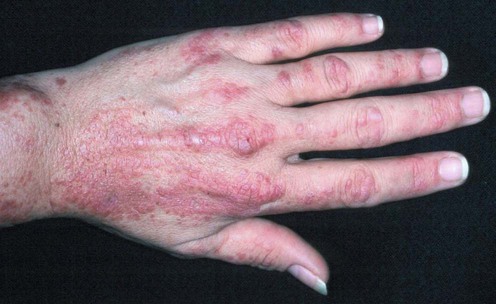
- Skin changes. A violet-colored or dusky red rash develops, most commonly on your face and eyelids and on knuckles, elbows, knees, chest and back. The rash, which can be itchy and painful, is often the first sign of dermatomyositis.
- Muscle weakness. Progressive muscle weakness involves the muscles closest to the trunk, such as those in your hips, thighs, shoulders, upper arms and neck. The weakness affects both the left and right sides of your body, and tends to gradually worsen.
When to see a doctor
Seek medical attention if you develop muscle weakness or an unexplained rash.
Causes
The cause of dermatomyositis is unknown, but the disease has much in common with autoimmune disorders, in which your immune system mistakenly attacks your body tissues.
Small blood vessels in muscular tissue are particularly affected in dermatomyositis. Inflammatory cells surround the blood vessels and eventually lead to destruction of muscle fibers.
Complications
Possible complications of dermatomyositis include:
- Difficulty swallowing. If the muscles in your esophagus are affected, you can have problems swallowing (dysphagia), which can cause weight loss and malnutrition.
- Aspiration pneumonia. Difficulty swallowing can also cause you to breathe food or liquids, including saliva, into your lungs (aspiration).
- Breathing problems. If the condition affects your chest muscles, you might have breathing problems, such as shortness of breath.
- Calcium deposits. These can occur in your muscles, skin and connective tissues (calcinosis) as the disease progresses. These deposits are more common in children with dermatomyositis and develop earlier in the course of the disease.
Associated conditions
Dermatomyositis may cause other conditions or put you at higher risk of developing them, including:
- Raynaud's phenomenon. This condition causes your fingers, toes, cheeks, nose and ears to turn pale when exposed to cold temperatures.
- Other connective tissue diseases. Other conditions, such as lupus, rheumatoid arthritis, scleroderma and Sjogren's syndrome, can occur with dermatomyositis (overlap syndromes).
- Cardiovascular disease. Dermatomyositis can cause heart muscle inflammation (myocarditis). In a small number of people who have dermatomyositis, congestive heart failure and heart arrhythmias develop.
- Lung disease. Interstitial lung disease can occur with dermatomyositis. Interstitial lung disease refers to a group of disorders that cause scarring (fibrosis) of lung tissue, making the lungs stiff and inelastic. Signs and symptoms include a dry cough and shortness of breath.
- Cancer. Dermatomyositis in adults has been linked to an increased likelihood of developing cancer, particularly of the cervix, lungs, pancreas, breasts, ovaries and gastrointestinal tract. Risk of cancer increases with age, although it appears to level off three years or so after a diagnosis of dermatomyositis. Dermatomyositis can also develop after you receive a diagnosis of cancer.
-
Dermatomyositis - An Overview
- 1. Dermatomyositis - An Overview Dermatomyositis is an uncommon inflammatory disease characterized by the muscle weakness and skin rash. This kind of disease is more common in adults and children alike. This disease generally occurs in the age group of 40 years to early 60’s. In children, the disease affects between 5 to 15 years of age. This kind of disease is more common in females as compared to males. There is no particular remedy for this disease, but periods of remission happens when symptoms improve gradually. Treatment can clear the skin rash and helps to regain muscle strength and functioning of the muscles. Dermatomyositis causes The exact cause of this disease is still not clear. However, researchers have found similarities between this disease and auto-immune disorders. Doctors have also established the link between these two diseases. Auto-immune disease occurs when the body’s disease fighting cells also known as anti-bodies attacks healthy body cells. It leads to compromised immune system. Viral infection may also contribute to this disease. Symptoms of disease The major symptoms of this disease are mentioned hereunder: Skin changes- When a person suffers from this disease a violet coloured red rash appears on the skin and most commonly on your face and eyelids and areas around nails, knuckles, back and chest. The rash can become patchy with bluish colour discoloration and is often labelled as Dermatomyositis. Weakness in muscle- As the disease progresses, muscle weakness appears like the muscles close to the trunk, upper arms, and back gets affected. The weakness affects both the right and left sides of the body and it gradually worsens with time. Other symptoms of this disease include: Fatigue Problem in swallowing Fever Hard calcium deposits under the skin surface Fatigue Weight Loss Fever Diagnosis Dermatomyositis is generally considered as inflammatory disease that is easy to diagnose because rash is associated with it. Your doctor will be able to diagnose the disease with amyopathic Dermatomyositis if you don’t experience muscle weakness. Other tests that needs to be performed to confirm it includes: Magnetic Resource Imaging- to have a look at abnormal muscles Electromyography- to record electrical impulses to control the muscles Blood Analysis- to check the level of anti-bodies Muscle Biopsy-sample muscle will show inflammation and other problems related to the disease
- 2. Skin Biopsy-skin sample will show the changes on the skin Dermatomyositis treatment Though there is no remedy for this disease, but treatment can improve the condition of the skin and muscle weakness. Prompt treatment improves the condition Corticosteroid medication- In majority of the cases, corticosteroid medications are the most preferred way of treating the disease. It can be ingested orally or can be applied on the skin. The medicine lowers the response of the immune system, which ultimately reduces inflammation causing antibodies. The doctor will increase the dose of corticosteroids and then suddenly lower after few weeks. In most cases, the medications like prednisone are the preferred way of treatment. Medicines that are used to control the side effects of corticosteroids like Azasan and methotrexate may be used in case you have further complications. IVIG- IVIG uses healthy antibodies to block the attacking anti-bodies that target your muscles and skin. IVIG medication includes a mixture of antibodies that have been collected from several healthy individuals, who donate their blood. The treatments don’t last for a long tenure, and it is likely you need infusion of six to eight weeks. Thus, Dermatomyositis is a disorder that can cause serious damage and may lead to medical complications if not treated promptly.
My report on my thighs, it has never been the same....STUDY: MRI LOWER EXTREMITY RIGHT THIGH WITH AND WITHOUT CONTRAST REASON FOR EXAM: Female, 42 years old. Dermatomyositis TECHNIQUE: Standardized fat and water weighted pulse sequences were obtained in all 3 orthogonal planes, post contrast administration. 13 ml of Multihance contrast material was administered intravenously for the contrast portion of the examination. COMPARISON: None. ___________________________________ FINDINGS: There is extensive edema with mild enhancement of the muscles of the thigh, involving all compartments (image 4, 5, 11, 18, 25, 34/41, 1 10, 20, 29/37 axial inversion recovery, 33, 24, 20/41 axial T1 fat-sat postcontrast, 13/37 axial T1 fat-sat postcontrast). Myofascial involvement is present. No skin involvement is demonstrated. There is no soft tissue mass. There is no fluid collection. There is no hematoma. The right femur demonstrates no fracture, infiltrative marrow process or osteonecrosis. ___________________________________ IMPRESSION: Extensive myositis, right thigh STUDY: MRI LOWER EXTREMITY LEFT THIGH WITH AND WITHOUT CONTRAST REASON FOR EXAM: Female, 42 years old. Dermatomyositis TECHNIQUE: Standardized fat and water weighted pulse sequences were obtained in all 3 orthogonal planes, post contrast administration. 13 ml of Multihance contrast material was administered intravenously for the contrast portion of the examination. COMPARISON: None. ___________________________________ FINDINGS: There is extensive edema with mild enhancement of the muscles of the thigh, involving all compartments (image 4, 5, 11, 18, 25, 34/41, 1 10, 20, 29/37 axial inversion recovery, 33, 24, 20/41 axial T1 fat-sat postcontrast, 13/37 axial T1 fat-sat postcontrast). There is myofascial involvement. There is no skin involvement. There is no soft tissue mass. There is no fluid collection. There is no hematoma. The right femur demonstrates no fracture, infiltrative marrow process or osteonecrosis. ___________________________________ IMPRESSION:
No comments:
Post a Comment